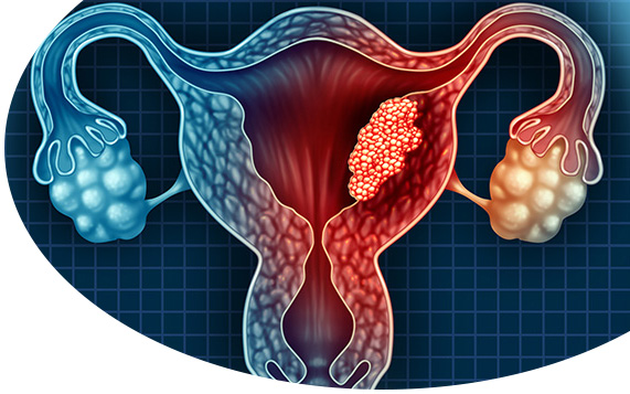
The condition of uncontrolled proliferation of cells in the layers of the uterus is called “uterine cancer“.
Uterus is consisted of two parts, the body of the uterus and the cervix. The body of the uterus has three layers:
- There is the membrane called “endometrium” in the inner layer of the uterus.
- The middle layer of the uterus consists of a thick layer of muscle called “myometrium“.
- The outer layer of the uterus contains the outer membrane of the uterus called “serosa”.
Approximately 95% of uterine cancers occur in epithelial and glandular tissues, which are called “carcinoma“. When the uterine cancer occurs in the endometrium, i.e. intrauterine membrane, these cancers are defined as “endometrial carcinoma“.
Another type of cancer that starts in the uterus is carcinosarcoma, and these cancers features both sarcoma (onset of cancer in muscle tissue) and carcinokama.
Types of Uterine Cancer
- Uterine sarcoma: It occurs at the muscle layer called myometrium in the uterus.
- Endometrial Carcinoma: It is the type of cancer that begins in the inner layer of the uterus.
Symptoms of Uterine Cancer
Abnormal vaginal bleeding is is the most common one of the uterine cancer symptoms. In particular, post-menopausal vaginal bleeding is followed by bleeding between the menstrual cycles and vaginal discharge.
The symptoms of uterine cancer may vary from person to person and depend on the stage of the disease. The symptoms of the people with uterine cancer and their severity may be different even at the same stage.
Causes of Uterine Cancer
The causes of uterine cancer are not clearly known, but the presence of factors increasing the risk of uterine cancer is accepted. Increased age, hormonal change, diabetes, previous endometrial hyperplasia and ovarian cancer, malnutrition, genetic predisposition, and early onset of the menstrual cycle may be examples of factors that increase the risk of developing uterine cancer.
Diagnosis of Uterine Cancer
For the diagnosis of endometrial cancer, the history of the patient should be initially heard and physical examination should be performed. The uterus, ovaries and fallopian tubes are then viewed with ultrasound. Pelvic ultrasound or transvaginal ultrasound may be required at this stage. Endometrial biopsy is performed to distinguish endometrial hyperplasia or uterine cancer as a result of suspicious findings. Endometrial biopsy provides accurate diagnosis of uterine cancer. Except for the biopsy, the diagnosis is clarified by hysteroscopy, dilatation and curettage.
Treatment of Uterine Cancer
Treatment of uterine cancer, as with all other types of cancer, is planned according to the stage of the disease and the general health of the patient. If the cancerous cells are not yet spread to the surrounding organs, the uterus can be removed with hysterectomy. Lymph nodules, ovaries, and fallopian tubes can also be removed from the body during hysterectomy.
Radiotherapy, chemotherapy and hormone therapy can be applied with or without hysterectomy performed by laparoscopic method in the treatment of uterine cancer. The gynecological oncologist will determine the treatment method that may give the best results.
We, as KADOMER, mobilize all the possibilities of technology for the diagnosis of disease as well as our experience for the success of the treatment processes.